
Salvatore Vendemmia - Aversa, Ilaria Pezone - Aversa, Maria Vendemmia - Naples
INTRODUCTION
An interesting work of Prof. Pasquale D'Acunzo, performed in the United States, in the laboratory of Professor Efrat Levy, in the year 2021, reveals the presence of a new subtype of extracellular vesicles of mitochondrial origin, altered in vivo and in vitro, due to aging or other pathological causes. The conclusions of this excellent research make us know and understand that there are connections between the presence and quantity of these new microvesicles with Down syndrome and other mitochondrial dysfunctions in independent sex mode.
NATURE AND RELEASE OF EXTRACELLULAR MICROVESICLES
Nanoscopic vesicles ( evs ) released by cells of extracellular fluid are secreted by all cells studied, and also by prokaryotes and plant cells. They originate from the plasma membrane, have a magnitude of 100-1000 nm, and are also called ECTOSOMES. The ESOSOMI, on the other hand, are produced by the endocytic system as endoluminal vesicles within vesicular bodies (MVB), have a magnitude of 50-200 nm and are released outside when the MVB merges with the plasma membrane. Extracellular microvesicles (evs) are fundamental for pathology and brain fisiology because they allow intercellular communication, eliminate indigestible toxic material inside the lysosome and regulate the immune system, flogosi and gene expression (myrrh).
FIG. N. 1
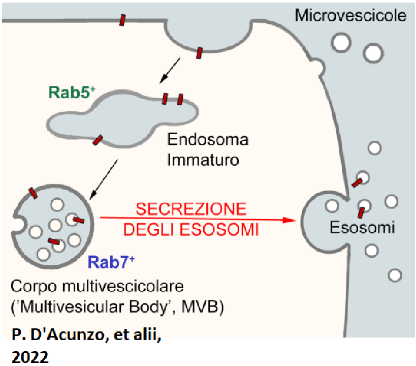
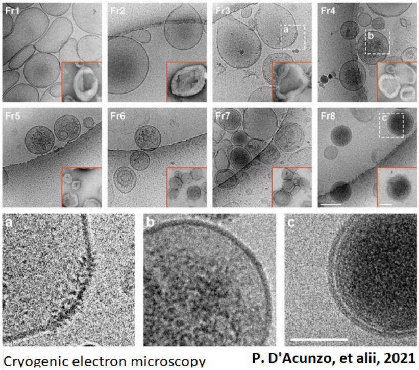
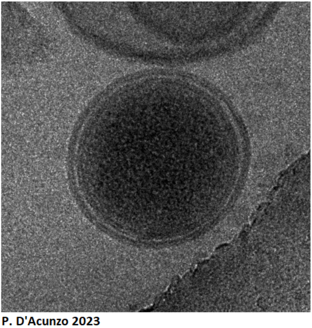
The evs can be isolated from biological liquids and have proved useful to provide diagnostic information. They can be used as vectors to encapsulate the drugs. Liposomes have been used mainly in vaccines for COVID 19 of Modern and Pfizer (giving rise to artificial evs). Research has shown that evs isolated from murine brains contain proteins mitochondrial. Accurate biochemical analyses suggest that in the brain NOT THERE ARE ONLY TWO SUBTYPES OF EVS, BUT THREE SUBTYPES. Therefore this new subtype was named evs MITOVESCICOLE. Finally it is important to know that mitovesciculums are evs with double membrane and an internal matrix very rich in electrons.
FIG: N. 2
WHAT ARE MITOVESCICULA , HOW ARE THEY GENERATED ?
It is probably " mini-mitochondria " very similar to those that have them originated and able to produce ATP. Certainly they represent a different group from the groups already characterized: microvesicles and exosomes. They are in space extracellular brain both in vivo and in the medium of in vitro culture cells. They contain a selective subgroup of mitochondrial proteins and have, in vitro, a metabolism because under appropriate conditions they produce ATP and maintain the enzymatic activity of mitovescicular proteins MAO-A and MAO-B. Mitochondrial damage stimulates the release of mitovescicula. Research has shown mitochondrial neurons produce double membrane vesicles when exposed to ROS.
FIG N 3.
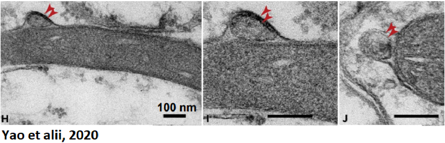
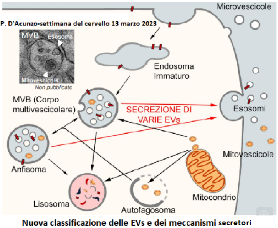
Neuronal mitochondria on different animals (rats, mice, drosophil, planar) produce vesicles with characteristics similar to mitovescicula: double membrane; size. They are produced in every neuronal region: cell body, dendrites, synaptic terminals. All this leads us to believe that we have learned of a new and interesting classification of extracellular vesicles and their mechanisms of secretion.
FIG N 4
MITOCHONDRIAL AND MITOCHONDRIAL DAMAGE
Mitochondrial damage in vitro and also in vivo shows altered biology mitovescicular. It now seems evident that in the presence of mitochondrial dysfunctions (ageing , Down’s syndrome , cocaine intake ,etc ) their alteration.
FIG. N.5
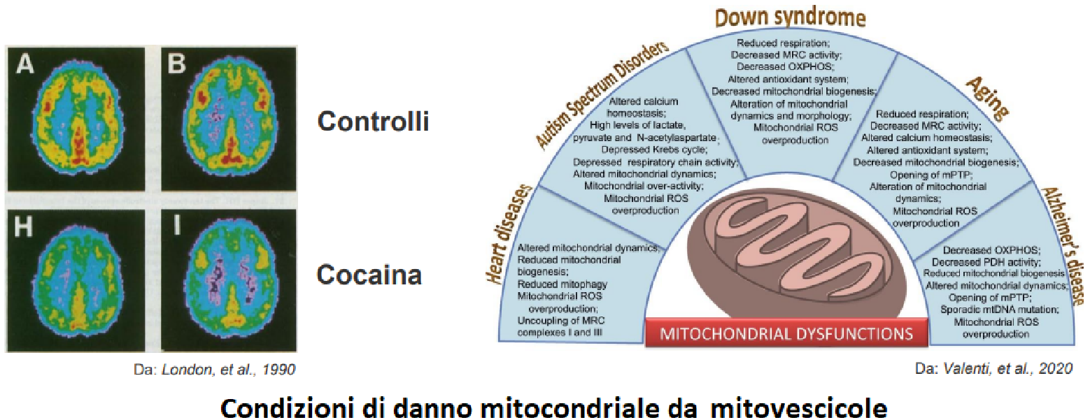
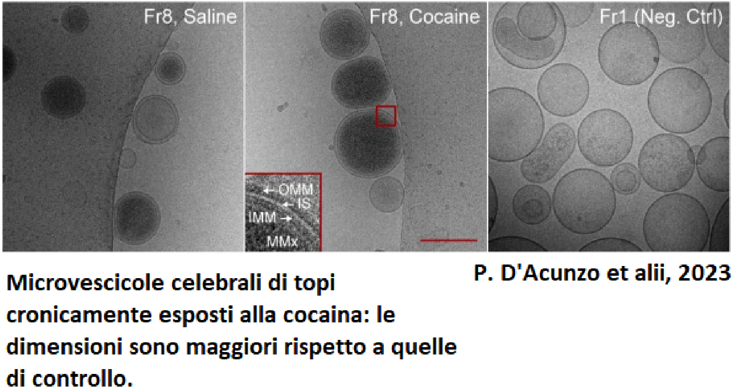
In Down syndrome the levels of mitovesciculum are increased, as well as in other mitochondrial dysfunctions tested, in an independent sex. It has also been established that not only the secretion, but also the composition and the mitovescicular dimension is altered in pathological conditions. Cocaine alters the content of cerebral mitovescicula and its effetto on the functionality and composition of mitovescicula is sex-dependent. In fact, careful research on mice has shown that protein alterations of mitovescicula are evident only in male mice exposed to cocaine, and not in women . ATP production is reduced only in male mice and not in females. In both sexes, the production of MAO-A (the protein involved in dopamine metabolism) is reduced.
FIG. N.6
CONCLUSIONS
In the brain there are THREE subtypes of EVs and not two!
Mitovesciculums are a group of newly identified extracellular vesicles of mitochondrial origin, different from the two groups already known (microvesicles and exosomes). They are located in the extracellular space of the brain both in vitro and in vivo .
They have unique characteristics compared to microvesicles and exosomes (double membrane, increased density, cardiolipin, etc.). Also if we compared them to mitochondria would have smaller dimensions, absence of most proteins mitochondrial, no crests, etc. Any mitochondrial dysfunction stimulates its release, both in vitro and in vivo. Their contents are altered in non-physiological conditions.
Unfortunately have not yet been well explored any roles in regulating stress oxidative, synaptic regulation, neuroinflammation and homeostasis cerebral.
Knowing this suggestive and extraordinary topic is very useful for our culture and education, but we hope that it will be an incentive for further and useful research that will certainly help our future generations.
FIG. N.7
BIBLIOGRAPHY
2023. D'Acunzo- Mitovescicole: a new subtype of extrscellular vesicles of mitochondrial origin altered in vivo with aging and pathology. Dep.of Psychiatry, New York University, Grossman School of Medicine - USA Brain Week , 13 March 2023.
Thanks to young student Aldo Russo for the help he made in filling out the work